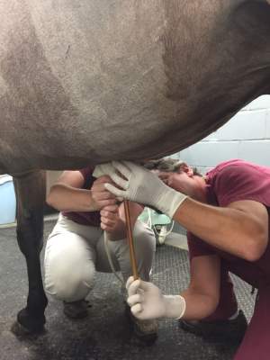
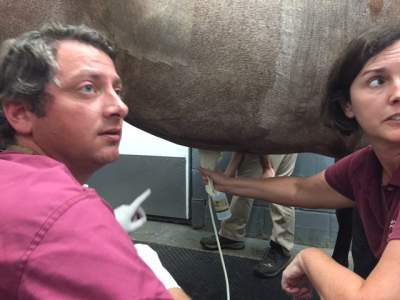
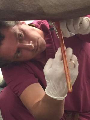
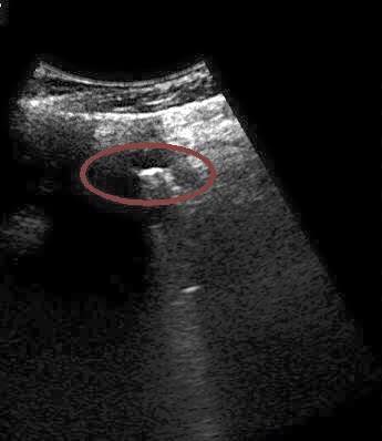
 An ultrasound is an invaluable tool. It gives us x-ray vision. In this case, it is helping Dr. Rich Hartman guide a tube into proper position to drain fluid.
The drain was placed in the horse to treat peritonitis, inflammation / infection of the abdominal cavity. By placing a tube in the horse’s abdomen it allowed Dr. Hartman and Dr. Dawn Hoover to drain the infected fluid and flush the abdomen with sterile fluids.
More About The Ultrasound Image:
The first thing to understand is that the ultrasound picture is upside down. The top of the image is the bottom of the horse’s belly. The bright white area in the red circle is the end of the metal trocar, which we use to introduce the tube into the horse’s abdomen. The ultrasound cannot penetrate or see through the metal, resulting in the white shadowing beyond the end of the trocar.
